Personalized Medical Care & Dedicated International Patient Concierge

Diagnosis & Evaluation · Needs Clinic Confirmation
Vascular Ultrasound Examination
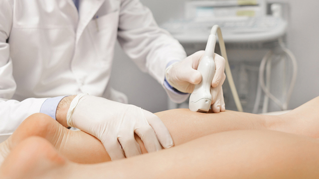
A vascular ultrasound examination evaluates the condition of the leg veins — deep, saphenous, subcutaneous and perforating — assessing their size, shape, location and blood-flow patterns, and showing reflux in real time.
Schedule a ConsultationVascular Ultrasound Examination
Medically reviewed by Dr. Dong-Hyuk Kim, Board-Certified General Surgeon, Medical DirectorIum Hazi Varicose Vein Clinic, Seocho-gu, Seoul
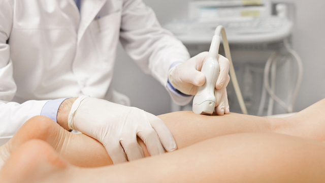
A vascular ultrasound examination is the imaging step inside a varicose vein evaluation that shows what the leg veins are actually doing. It assesses the deep veins, the saphenous veins, the subcutaneous veins and the perforating veins, records their size, shape and location, and shows reflux — blood moving the wrong way — as it happens. At this clinic it is performed as part of assessment and treatment planning rather than offered as a standalone service.
What Is a Vascular Ultrasound Examination?
A vascular ultrasound examination evaluates the condition of the leg veins — deep, saphenous, subcutaneous and perforating — assessing their size, shape, location and blood-flow patterns, and showing reflux in real time.
It is non-invasive: nothing is injected and nothing enters the body.
It looks at four groups of veins together, not only the vein that is visible from the outside.
It measures structure and movement: what the veins look like, and which way blood travels through them.
Reflux is demonstrated dynamically, as flow, rather than inferred from a still picture.
Its output is the finding set a treatment plan is built on.
The reason movement matters is the mechanism of the disease. The American Venous Forum describes leg veins as depending on one-way valves and the calf muscle pump, with backward flow — venous reflux — following when valves fail. MedlinePlus, from the US National Library of Medicine, describes duplex ultrasound generally as combining grey-scale imaging with Doppler to show how blood moves through arteries and veins, less invasive than venography and listed as carrying no risks. For chronic venous insufficiency specifically, the Society for Vascular Surgery describes diagnosis as resting on history and physical examination with referral for duplex ultrasound to evaluate blood flow and vein structure.
Who Performs the Vascular Ultrasound Examination at Ium Hazi Varicose Vein Clinic
Dr. Dong-Hyuk Kim
Board-certified general surgeon
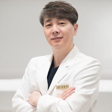
Dr. Dong-Hyuk Kim is a board-certified general surgeon and the Medical Director of Ium Hazi Varicose Vein Clinic. He completed his medical education at Sungkyunkwan University School of Medicine and serves as Executive Director of the Korean Society of Phlebology.
Ium Hazi Varicose Vein Clinic operates a single-director system: Dr. Dong-Hyuk Kim personally performs every consultation, diagnostic ultrasound, procedure and post-operative follow-up, and the clinic deliberately limits its schedule to one or two surgical cases per day.
Who holds the probe is not an administrative detail here. Venous ultrasound image quality depends heavily on the skill of the person performing the scan, as RadiologyInfo.org from the Radiological Society of North America and the American College of Radiology notes for the examination category, and the same source records that the technique uses no ionizing radiation and no contrast agent. Vascular ultrasound diagnosis and evaluation is one of Dr. Kim's documented core specializations, and his documented teaching activity includes serving as a tutor on a varicose vein ultrasonography hands-on course at the Spring Conference of the Society of Surgical Ultrasound in 2014.
This page does not cite case counts, awards, ratings or patient testimonials. Where the clinic publishes such figures they are self-reported and unaudited, and they do not describe what any individual patient should expect.
Who Is a Good Candidate for a Vascular Ultrasound Examination?
People with suspected varicose veins who need the superficial and deep veins of the legs evaluated.
People concerned about abnormal venous blood flow or reflux.
Adults with leg swelling, pain, visible veins or suspected blood-flow problems that call for assessment.
Patients who need the affected veins identified before a treatment plan can be made.
There are generally no major contraindications, because the examination is non-invasive. The clinic documents one practical limit: the examination may need to be modified or postponed when the area cannot be adequately accessed.
One point belongs here rather than in the small print. This examination is part of assessment and treatment planning at this clinic, not a service booked on its own for its own sake; the clinic's own content rules exclude a standalone simple vascular ultrasound examination from promotion. If what you want is the finding — what is wrong with these veins and what follows from it — that is a consultation, and the ultrasound is part of it.
What to Expect: Procedure, Recovery, and Results Timeline
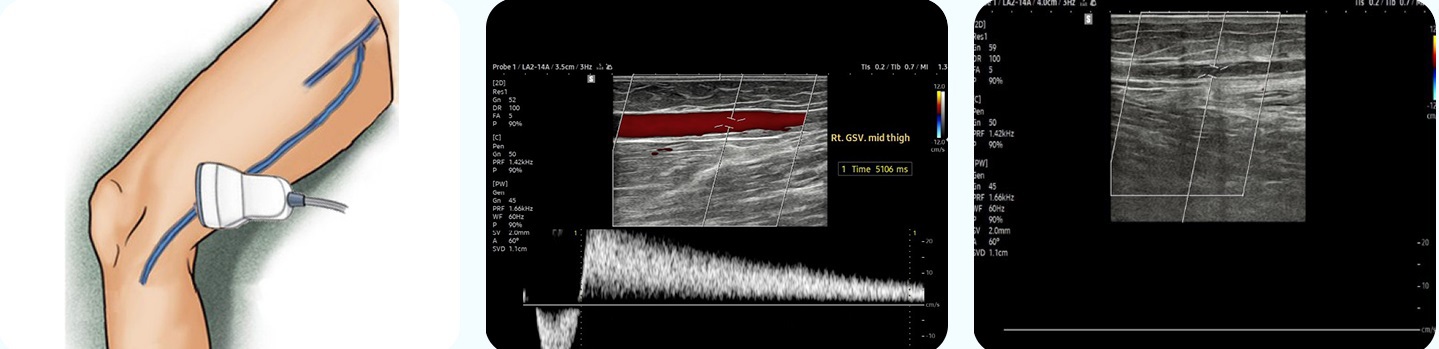
The clinic documents the sequence as: the examination area is prepared for ultrasound; a probe is used to examine the veins of the legs; the deep, saphenous, subcutaneous and perforating veins are assessed; blood flow and reflux are observed in real time; the findings are explained using both the ultrasound images and the dynamic reflux flow; and, if requested, the images are saved directly to a USB drive.
Stage | What happens | Timing |
|---|---|---|
Preparation | Usually none required for a leg vascular ultrasound; follow any clinic instructions given | Before the visit |
Scanning | Probe assessment of the deep, saphenous, subcutaneous and perforating veins | Same visit; duration depends on how many veins and areas are assessed |
Reflux assessment | Reflux examined in the inguinal, thigh, popliteal and calf regions | During the scan |
Explanation | Findings shown as static images and as moving reflux flow | As they are seen |
Records | Images saved to a USB drive on request | End of the examination |
Afterwards | Normal activities generally resumed immediately | Immediate |
Anaesthesia is not required. Most patients feel little or nothing; some notice mild pressure or brief discomfort as the probe passes over a tender area. No specific recovery period is needed and no special aftercare applies, unless another procedure is performed at the same visit.
Results are not posted out later. Because the examination is interpreted as it is performed, the venous condition and the reflux flow are explained during the scan, and the discussion of what follows happens in the same visit. For international patients, a same-day clinic visit is generally sufficient for the examination itself; allow more time if consultation, further testing or treatment is planned. After a diagnostic ultrasound alone, travel is generally possible once the examination and consultation are complete.
What the Four Vein Groups Mean
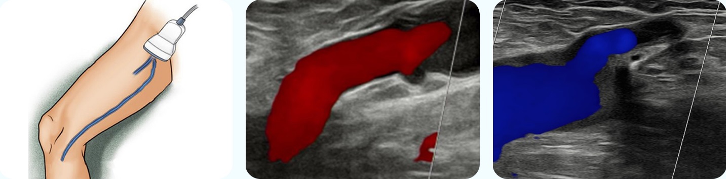
An examination that only looked at the bulging vein would answer the wrong question. The clinic documents assessment of four groups, and each answers something different.
Vein group | Why it is examined |
|---|---|
Deep veins | The main drainage route of the leg; their condition affects whether and how superficial veins can be treated |
Saphenous veins | The great and small saphenous trunks, the usual site of the reflux that drives visible varicose veins |
Subcutaneous veins | The surface veins, including the ones a patient can see and feel |
Perforating veins | The connections between the superficial and deep systems, where reflux can cross between them |
The clinic also documents where reflux itself is assessed — the inguinal, thigh, popliteal and calf regions — and that the patient's condition is explained using both static ultrasound images and dynamic, real-time reflux flow rather than images alone. The equipment used provides static vascular images and real-time visualisation of reflux blood flow, with a system for saving examination images to a USB drive when requested.
The clinic's documented equipment includes a Samsung Medison V8 high-resolution vascular ultrasound system, a VeinViewer Flex vein visualisation device, a 4th-generation 1940 nm endovenous laser system and a Medtronic ClosureRFG and RFG 3D radiofrequency system. Instruments are sterilised by autoclave and ethylene oxide gas, and single-use devices are not reused.
Regulatory approval, clearance or certification status for the devices used at this clinic is not documented in the clinic's source material, so no approval, clearance or certification claim is made on this page.
Combining the Vascular Ultrasound Examination with Other Treatments
The examination is diagnostic rather than therapeutic, so it does not combine with treatment so much as precede and follow it. The clinic documents that where venous reflux or varicose veins are identified, the vascular ultrasound is performed as part of the diagnostic evaluation before or alongside appropriate varicose vein treatment, with the specific combination depending on the findings.
Diagnosis & Evaluation — the consultation and assessment this examination sits inside.
Endovenous Vein Ablation — where a refluxing trunk is identified, the examination establishes which one and guides the treatment itself.
Micro-Phlebectomy — where prominent surface veins are to be removed, the examination maps them.
Post-treatment examinations, which the clinic includes in aftercare and which allow blood-flow findings to be compared before and after treatment.
Depending on the findings, further diagnostic assessment or treatment may be considered by the physician; the examination itself does not decide that.
Safety, Precision, and Clinic Standards
The risks below are the ones this clinic documents in its own treatment information. No procedure is free of risk, and a written list is not a substitute for the discussion of risks and alternatives that takes place during consultation.
A vascular ultrasound examination is generally considered a safe, non-invasive examination, and significant complications are uncommon. The effect patients report is mild pressure or temporary discomfort from the probe. There is no radiation exposure and no meaningful downtime.
The documented risk-reduction measure is the thoroughness of the examination itself: assessing the deep, saphenous, subcutaneous and perforating veins, evaluating reflux with real-time ultrasound, and explaining the findings using both static images and dynamic blood-flow visualisation. The clinic's stated quality process is doctor-led diagnosis, consultation, surgery and post-operative care; pre-operative vascular ultrasound and clinical evaluation; a deliberately limited daily surgical caseload; strict sterilisation using autoclave and ethylene oxide gas systems; no reuse of single-use devices; and post-operative vascular ultrasound monitoring and follow-up care.
The clinic is a Registered Medical Institution for Attracting Foreign Patients, certified by the Republic of Korea's Ministry of Health and Welfare under the Medical Overseas Expansion and Foreign Patients Attraction Act.
This clinic does not publish closure rates, success rates or recurrence rates, so none are stated here. Figures published for a treatment category as a whole do not describe the results of any individual clinic or patient.
This page does not quote prices and does not describe insurance coverage. Fees, what a quotation includes, and any question of reimbursement are discussed during consultation, and patients outside Korea should confirm coverage with their own insurer.
The Bottom Line
The examination assesses four vein groups — deep, saphenous, subcutaneous and perforating — not only the vein you can see.
Reflux is shown as movement, in the inguinal, thigh, popliteal and calf regions, and explained while it is being seen.
It is non-invasive, needs no anaesthesia and no preparation, and normally requires no recovery.
Images can be saved to a USB drive on request, so the findings travel with you.
It belongs inside assessment and treatment planning at this clinic rather than being booked on its own.
Frequently Asked Questions
Does the scan hurt, and do I need to prepare?
The examination is generally painless and usually needs no special preparation. Some patients notice mild pressure or brief discomfort as the probe moves across the leg, particularly over an area that is already tender. No anaesthesia is used, because nothing invasive takes place.
Is there any radiation?
No. Venous ultrasound as a category uses sound waves, with no ionizing radiation and no contrast agent, which is part of why it is the routine test for this question rather than a last resort. That also means there is no cumulative exposure consideration if the examination is repeated at follow-up.
How long does it take?
It is generally completed within a single clinic visit, and the exact time depends on how many veins and how many areas are being assessed. A scan covering both legs and all four vein groups takes longer than one focused on a single region.
Can I have the examination on its own, without a consultation?
At this clinic the examination is part of assessment and treatment planning rather than a standalone service. That is the clinic's own rule, not a scheduling preference: an image without an interpretation and a plan does not answer the question most patients are actually asking.
What happens to the images afterwards?
They can be saved directly to a USB drive if you ask for that at the examination. The clinic documents this as an available option, which matters most for patients who will continue their care elsewhere and want the record of what was found.
Will the scan tell me which treatment I need?
It tells the doctor which veins are refluxing, where and how badly, and that is what the treatment decision is made from — but the decision itself comes from the consultation, weighing the findings against your symptoms, anatomy and medical history. The examination narrows the question; it does not answer it on its own.
What if the scan finds nothing wrong?
That is a result, not a wasted visit. Ruling out significant reflux changes what the symptoms are likely to be and what should be looked at next, and the clinic's stated approach is to recommend only the treatment a finding actually justifies rather than treating on appearance.
Book a Consultation at Ium Hazi Varicose Vein Clinic
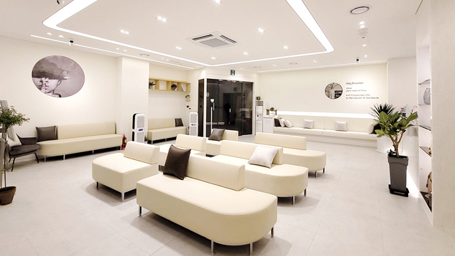
If you are considering a leg vein ultrasound in Seoul, book it as part of a varicose vein consultation, which is how the examination is used here.
To arrange a consultation, use the clinic's contact page or call . English-language support is available for international patients.
Ium Hazi Varicose Vein Clinic is on the 2nd floor of K-Tower, 320 Seocho-daero, Seocho-gu, Seoul 06633, Republic of Korea. More about the clinic is on its About Us page.
Disclaimer
Individual results may vary. All surgical and non-surgical procedures have potential risks and limitations. Your suitability for treatment, expected results, costs, and recovery time will be discussed with Dr. Dong-Hyuk Kim during your personal consultation. The information provided is for general educational purposes and does not guarantee a specific treatment outcome.
Sources & Further Reading
Other Services
Explore additional treatments offered by our specialists.
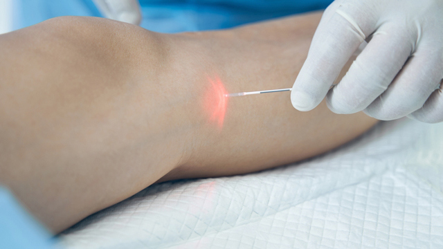
Endovenous Laser Ablation
Endovenous laser ablation is the closure of an abnormal vein from the inside using laser energy delivered along a fibre placed under ultrasound guidance, used for appropriately diagnosed venous reflux and the varicose vein symptoms that follow from it.
Learn More
Vascular Ultrasound Examination
A vascular ultrasound examination evaluates the condition of the leg veins — deep, saphenous, subcutaneous and perforating — assessing their size, shape, location and blood-flow patterns, and showing reflux in real time.
Learn More
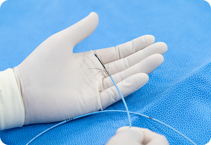
Mechanochemical Ablation
Mechanochemical ablation closes a refluxing vein by combining two actions in one catheter pass: mechanical irritation of the inner vein wall and delivery of a chemical sclerosing agent, without heat.
Learn More