Personalized Medical Care & Dedicated International Patient Concierge

Medical Guide
Untreated Varicose Veins: Blood Clot and Skin Ulcer Risk
Untreated varicose veins can be associated with thrombosis and skin ulcers. Here is what the evidence describes, how the risk varies, and when to be assessed.
Schedule a ConsultationUntreated Varicose Veins: Blood Clot and Skin Ulcer Risk
Medically reviewed by Dr. Dong-Hyuk Kim, Board-Certified General Surgeon, Medical Director of Ium Hazi Varicose Vein Clinic
Untreated varicose veins can be associated with blood clots and with skin ulcers, and Ium Hazi Varicose Vein Clinic states it directly. Its own list names four things varicose veins may be associated with — pain, swelling, thrombosis, and ulceration of the skin — and it adds that where the disease is untreated or progressing, circulation problems and, in more severe cases, complications of that kind can follow. What the statement does not say is that these outcomes are usual, or that they arrive on a schedule. This article sets out what the evidence describes about each complication, what makes one leg more likely than another to reach that point, and what an assessment can establish about your own.
What "Untreated" Means Here
"Untreated" is not a single decision made once. Most people with varicose veins are not choosing between a procedure and nothing at all; they are living with a condition that has degrees, and their situation changes over years rather than weeks.
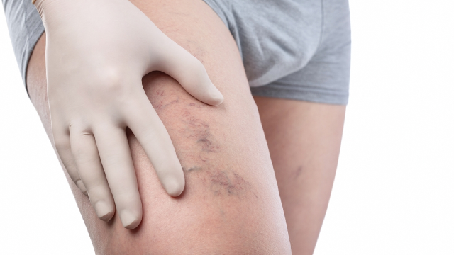
It is also worth separating two different things that both get described as untreated varicose veins. One is a leg with visible surface veins and no significant reflux underneath, where the veins are a cosmetic finding. The other is a leg where a saphenous trunk or a perforating vein is refluxing, blood is falling back down the leg with every standing minute, and the visible veins are the downstream expression of that. These two legs carry different outlooks, and they cannot be told apart by looking. Ium Hazi Varicose Vein Clinic uses vascular ultrasound rather than appearance to establish which is which.
That distinction governs everything below. The complications described here arise from sustained venous hypertension — persistently raised pressure inside the leg veins — rather than from the visible vein itself.
Superficial Vein Thrombosis and Deep Vein Thrombosis Are Not the Same Event
The word thrombosis covers two events with different implications, and conflating them makes the risk picture harder to read rather than easier.
A clot that forms inside a surface varicose vein produces a segment of vein that becomes firm, warm, red, and tender to touch, often over a few days, and the cord-like hardness can usually be felt. Ium Hazi Varicose Vein Clinic names inflammation of a superficial vein and superficial thrombophlebitis among the events that can occur in venous disease, and this is the complication most directly associated with the varicose vein itself: a dilated, slow-flowing vessel is a place where blood moves sluggishly. It is painful and it needs medical evaluation, both to manage it and because its relationship to the deep system has to be assessed.
A clot in a deep vein of the leg is a different matter. MedlinePlus, the patient information service of the US National Library of Medicine, describes deep vein thrombosis as a clot forming in a deep vein of the lower leg, the thigh, or the pelvis. Its stated signs are a leg that swells, hurts, feels tender to touch, feels warm, or shows red or discolored skin. Part of the clot can break away and travel to the lungs, where it becomes a pulmonary embolism, and that sudden breathlessness, chest pain that sharpens on breathing in, a rapid heartbeat, or coughing up blood are signs of that. Those are emergency symptoms and call for emergency care. The National Heart, Lung, and Blood Institute (NHLBI) treats deep vein thrombosis and pulmonary embolism together as venous thromboembolism for this reason.
The honest summary is that varicose veins sit in the same risk territory as deep vein thrombosis without being a straightforward cause of it. The Centers for Disease Control and Prevention (CDC) sets out the factors that raise venous thromboembolism risk, and the list is dominated by immobility, recent surgery or injury, cancer and its treatment, pregnancy and the period after it, older age, hormone medications, and a personal or family history of clots. Venous disease of the leg belongs in that picture, and a superficial clot in a varicose vein is one of the findings that prompts a clinician to look at the deep veins as well.
How Skin Damage Develops, and Why It Is Gradual
Skin ulceration is the other complication the clinic names, and it is the end of a long sequence rather than a sudden event.
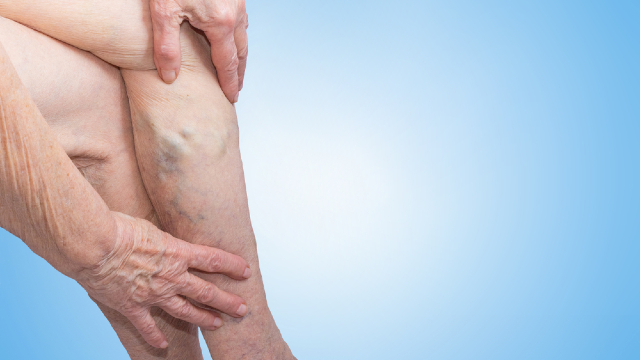
The driver is pressure. When reflux keeps venous pressure in the lower leg high through every standing hour, fluid and blood components are gradually forced out of the smallest vessels into the surrounding tissue. What follows is a recognizable progression: ankle swelling that no longer fully settles overnight; brown or rust-colored staining of the skin as iron from broken-down red cells is deposited; itching and eczema-like irritation returning to the same patch; and, over longer periods, tissue that becomes firm and inelastic. Skin in that state has a reduced tolerance for minor injury, and a break in it can be slow to close because the pressure that damaged it has not changed.
MedlinePlus includes sores and ulcers on the leg among the more serious consequences varicose veins can lead to, and the NHLBI likewise describes skin changes and sores as complications that can develop where venous disease is not addressed. What the evidence describes is a sequence with visible intermediate stages, which is the practically useful part: the staining, the persistent swelling, and the hardening are observable long before anything opens, and they are the findings that tell a clinician where a leg sits on that path. The management of an ulcer once it has formed is a separate subject involving wound care alongside the vein.
Bleeding, and Other Events Worth Naming
Two further events belong on an honest list.
A varicose vein lying just under the skin can bleed, sometimes after minor contact, and sometimes more than the size of the vein would suggest, because the pressure inside it is raised. Ium Hazi Varicose Vein Clinic lists bleeding among the possible problems in venous disease. The immediate response is to lie down, elevate the leg, and apply firm direct pressure, and then to be seen by a physician. A vein that has bled is a vein under pressure, and the pressure has a source that has not been identified yet.
The second is the general worsening of symptoms themselves. Ium Hazi Varicose Vein Clinic describes circulation problems, pain, and swelling as part of what untreated or progressing varicose veins can involve, and lists heaviness, cramps, and itching among the symptoms patients present with. This is not a complication in the dramatic sense, but for most people it is what actually changes, and it is the reason the clinic uses the effect on daily activities as its threshold for assessment rather than the presence of a named complication.
What Makes One Leg More Likely to Progress Than Another
Progression is not uniform, and the factors that separate one leg from another are largely the same ones that produced the varicose veins in the first place.
The NHLBI identifies family history, age, pregnancy, excess body weight, and long periods of standing or sitting among the factors associated with weakened or damaged vein valves. A previous deep vein clot deserves separate mention, because damage to a valve left behind by a past thrombosis can produce reflux that does not respond in the usual way. The extent of the reflux matters too: a refluxing saphenous trunk loads the lower leg differently from an isolated surface vein, which is exactly what ultrasound is used to distinguish.
What no set of risk factors can do is give an individual a timeline. The clinic's own framing is that suitability, findings, and outcomes are determined by examination in the individual case, and Dr. Dong-Hyuk Kim describes his approach as identifying the underlying cause accurately through precise vascular ultrasound and then providing only the treatment or combination of treatments that patient needs. A leg with skin staining and a refluxing trunk is in a different position from a leg with a few visible veins and normal flow, and the only way to know which one you have is to have it examined.
When to Have a Leg Assessed
Assessment is reasonable at the point where symptoms affect your daily activities, and it is appropriate sooner where specific findings are present. The clinic's own presenting list runs as follows: swelling, pain, a heavy feeling in the leg, itching, cramping, veins that are visible, and changes in the skin.
Several findings should be evaluated by a physician rather than watched: skin at the ankle that has darkened, thickened, or begun to itch repeatedly in the same place; a segment of vein that is firm, hot, red, and tender; any bleeding from a leg vein; and any open area on the lower leg. One leg that swells with pain, tenderness, or warmth, and any chest pain or breathlessness, belong in emergency care.
An assessment is also a reasonable step when nothing on that list applies and you simply want to know where you stand. A finding of no significant reflux is a real answer, not a wasted appointment, and it settles the question that the risk information above cannot settle from a distance.
What an Assessment Establishes
The examination answers three things: whether reflux is present, where it starts, and what state the tissue is in.
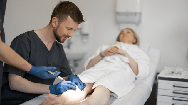
Ium Hazi Varicose Vein Clinic takes a medical history, examines the leg, and images the deep system, the saphenous veins, the subcutaneous veins, and the perforators by vascular ultrasound, logging the size, shape, and position of each vessel while flow and reflux are displayed as they happen. Nothing about the study is invasive and no anesthesia is needed. Sound waves, not ionizing radiation, form the picture in an ultrasound examination, a distinction the US Food and Drug Administration (FDA) sets out in its consumer material, and it is part of why imaging can be repeated over time to follow a leg rather than used once.
Where reflux is confirmed in a vein that can be safely treated, the clinic documents endovenous vein ablation as closing an abnormal vein so that blood is redirected through healthier veins, and identifies candidates as patients with symptomatic varicose veins or venous insufficiency caused by reflux in an affected vein, established through vascular examination and ultrasound. It also documents surgical options including point micro-surgery and cryoablation for veins better suited to removal, and graduated compression for symptom management, noting that a garment controls symptoms during wear rather than repairing the venous fault itself. Which of these applies is a physician's decision after imaging. A diagnostic consultation is where that determination is made, and it can be arranged through the clinic's contact page.
Treatment Changes the Picture Without Removing Risk
It would be misleading to present treatment as the elimination of the risks described above, and the clinic does not present it that way.
Closing a refluxing vein removes one driver of raised venous pressure in that leg. It does not make the venous system new. The clinic says that veins not treated, and veins that become affected afterward, can give problems later on, with long-run results differing between patients. Skin that has been damaged over years does not return to its earlier condition because a vein has been closed, and follow-up remains part of the model rather than an optional extra; its stated pattern is a review at roughly one week after treatment and imaging at about one to two months.
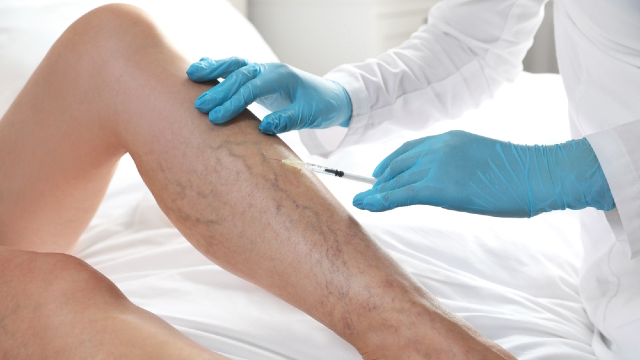
Procedures also carry their own risks, which do not shrink because the underlying situation is serious. For catheter-based closure the clinic lists bruising, swelling, tenderness, tightness, temporary numbness, skin irritation, and discomfort along the treated vein, and less commonly infection, nerve irritation, blood clots, skin injury, allergic reaction to materials used, and reopening of the treated vein. For injection-based treatment its list includes inflammation of a superficial vein, skin discoloration, injection-site irritation or ulceration, allergic reaction, incomplete treatment or recurrence, and, less commonly, clots including deep vein thrombosis. Skin ulceration appears there as a possible complication of treatment as well as of the disease, which is one reason method selection follows imaging rather than preference.
Who Would Carry Out the Assessment
A question about progression is a question about follow-up as much as about a single visit, and Ium Hazi Varicose Vein Clinic structures that explicitly: an examination roughly one week after treatment and imaging within about one to two months, with post-operative ultrasound monitoring recorded as a standing part of its process. The practice, in Seocho-gu, Seoul, has worked only on varicose veins since 2009, and one physician runs it. Dr. Dong-Hyuk Kim personally performs every consultation, every diagnostic ultrasound, and every procedure, so the same clinician who saw the leg at the start sees how it has changed.
Board certification in general surgery, medical training at Sungkyunkwan University School of Medicine, an Executive Director post with the Korean Society of Phlebology, membership of the Venous Disease Research Society, and an adjunct professorship at Samsung Medical Center make up his published profile. Experience is given as over 15 years confined to this condition. Its published statistic — 7,300+ saphenous vein surgeries, 2009 to 2024 — is the clinic's own count, and the practice received the Medical Asia 2024 Grand Prize in the varicose vein category.
Documented safety practice includes sterilization by autoclave and by E-O gas, disposable devices that are never reused, clinical evaluation with ultrasound before any operation, and a daily limit of one or two surgical cases. The about page describes the practice in more detail. If you are in the United States with a leg that already shows skin change or an open area, assessment by a physician where you are is the sensible first step.
The Bottom Line
- Ium Hazi Varicose Vein Clinic lists pain, swelling, thrombosis, and skin ulceration as possible associations of varicose veins, and describes circulation problems together with more severe complications where the disease goes untreated or continues to progress.
- A clot in a surface varicose vein and a clot in a deep vein are different events; the first produces a firm, hot, tender vein segment, and the second is described by MedlinePlus as deep vein thrombosis with the risk of a clot reaching the lungs.
- Skin ulceration develops at the end of a long sequence, with persistent ankle swelling, brown staining, itching, and hardened tissue appearing well before any break in the skin.
- Progression varies by individual, and the factors involved are largely those the NHLBI names for varicose veins themselves, including family history, age, pregnancy, weight, and prolonged standing or sitting.
- Treating a refluxing vein removes one driver of raised pressure; the clinic states that other veins can cause problems later, that long-term results vary, and that procedures carry their own risks.
Frequently Asked Questions
Do varicose veins directly cause deep vein thrombosis?
The relationship is one of shared territory rather than simple cause and effect. The CDC's list of factors raising venous thromboembolism risk is dominated by immobility, surgery, injury, cancer, pregnancy, age, hormone medications, and previous clots. A clot in a surface varicose vein is one of the findings that prompts a clinician to examine the deep veins as well, which is why it is assessed rather than managed at home.
How can I tell a superficial clot from a deep one?
A clot in a surface vein usually gives a firm, warm, red, tender cord you can feel along the vein. MedlinePlus describes deep vein thrombosis as producing swelling, pain, tenderness, warmth, and red or discolored skin in the leg, often without a palpable vein. Telling them apart reliably requires ultrasound, so both warrant medical assessment rather than self-diagnosis.
What are the earliest signs that my skin is being affected?
Ankle swelling that no longer settles fully overnight is usually first, followed by brown or rust-colored staining of the skin above the ankle and itching or eczema-like irritation that keeps returning to the same patch. Tissue that feels firmer and less elastic than the surrounding skin comes later. These changes appear before any break in the skin and are the findings worth showing a physician.
If my varicose veins do not hurt, do I still need an assessment?
Symptom severity and the amount of reflux underneath do not track each other reliably, which is why the clinic assesses venous findings by ultrasound rather than by appearance or discomfort. An assessment establishes whether a refluxing vein is present, and a result showing none is a useful answer. Whether treatment follows is a separate decision made on the findings.
Does wearing compression stockings prevent these complications?
Ium Hazi Varicose Vein Clinic's position is that a compression garment controls symptoms for as long as it is on the leg, without repairing the venous problem underneath and without stopping new vein problems from arising. It also says compression is not used in a leg with suspected or significant peripheral arterial disease until that has been assessed, and that a badly fitted garment can injure skin. It is a management tool used alongside assessment rather than instead of it.
Will treating the vein remove the risk of clots and ulcers?
No treatment can be presented that way. Closing a refluxing vein addresses one driver of raised venous pressure in that leg, but the clinic says a vein left alone, or one affected at a later point, may give problems afterward and that outcomes over the long run are not uniform. Procedures also carry their own listed risks, including blood clots and, for injection-based treatment, skin ulceration.
This article is for general informational purposes and does not constitute individualized medical advice. Candidacy, technique, and expected outcomes can only be determined through a consultation with a qualified physician, and this article is not a self-diagnosis tool.
References
- MedlinePlus: Varicose Veins
- MedlinePlus: Deep Vein Thrombosis
- MedlinePlus: Blood Clots
- NHLBI: Varicose Veins
- NHLBI: Venous Thromboembolism
- CDC: Risk Factors for Blood Clots
- CDC: About Venous Thromboembolism
- FDA: Ultrasound Imaging
More Articles
Continue reading our clinical guides.
Will My Varicose Veins Come Back After Treatment?
A closed vein and a new varicose vein are different events. Here is what recurrence after vein treatment in Korea means and how follow-up imaging detects it.
Learn More

What to Pack Before Flying to Korea for Vein Surgery
What to pack before flying to South Korea for vein surgery: documents, medication and supplement lists, clothing, compression, and what to arrange at home.
Learn More
What Are Varicose Veins? Causes, Symptoms, and Signs
Varicose veins are enlarged, twisted leg veins caused by valve dysfunction. Learn the causes, the symptoms they produce, and when to see a vein specialist.
Learn More