Personalized Medical Care & Dedicated International Patient Concierge

Medical Guide
Spider Veins vs. Varicose Veins vs. Reticular Veins
Spider, reticular, and varicose veins differ by size, depth, and what sits behind them. Here is how each is identified and why the label changes the plan.
Schedule a ConsultationSpider Veins vs. Varicose Veins vs. Reticular Veins: Understanding the Differences
Medically reviewed by Dr. Dong-Hyuk Kim, Board-Certified General Surgeon, Medical Director of Ium Hazi Varicose Vein Clinic
Spider veins, reticular veins, and varicose veins are three different sizes of vessel sitting at three different depths in the leg, and the words are not interchangeable even though patients and marketing pages often use them as if they were. Getting the label right matters because the label is the first step toward the question that actually decides treatment — what is feeding that vein. This article sets out how each type is identified, how the naming system a clinician uses differs from the one a patient uses, and why classification changes what happens next.
Two Different Naming Systems Are in Use at Once
Most of the confusion comes from a fact nobody states out loud: there are two parallel vocabularies for leg veins, and they answer different questions.
The first vocabulary describes what a vein looks like from outside — its caliber, its color through the skin, and whether it stands proud of the surface. Spider, reticular, and varicose are terms from this system. They are descriptive, they are what a patient can apply in front of a mirror, and they say nothing about cause.
The second vocabulary describes what a vein is anatomically and what job it does. A vascular examination works in this system, assessing deep veins, saphenous trunks, subcutaneous veins and perforators — the connecting vessels that run between the superficial and deep networks through the muscle layer. Ium Hazi Varicose Vein Clinic documents its ultrasound assessment in exactly these terms, evaluating the size, shape, and location of each group along with blood flow.
Neither vocabulary is wrong. But a description of appearance cannot be converted into a diagnosis, and this is the single most useful thing to understand before reading anything else about vein types.
Spider Veins
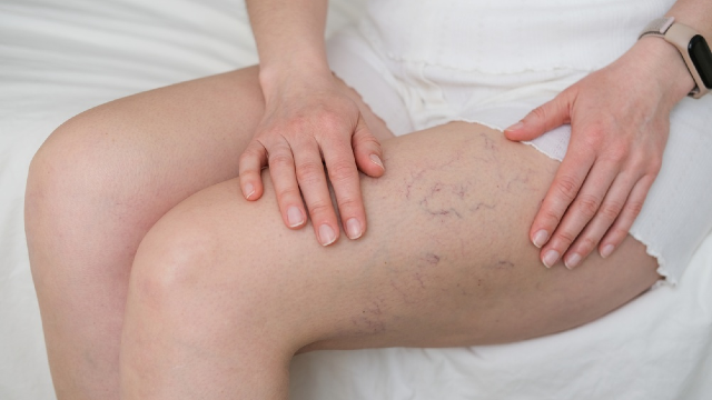
Spider veins are the smallest visible vessels, lying in the most superficial layer of the skin itself. They appear as fine red, purple, or blue lines, often branching outward from a point or spreading in a web or fan pattern, and they are flat — you can see them but you cannot feel them under a fingertip. The clinical term for these tiny surface vessels is telangiectasias.
They are common on the thighs, the backs of the calves, around the ankles, and on the face. Ium Hazi Varicose Vein Clinic describes cosmetic vein treatment as targeting visible spider veins and telangiectasias, and notes that facial telangiectasias are treated as well as leg vessels depending on the technique.
Spider veins on their own usually produce no symptoms at all. Where a patient does report something, the clinic describes it as localized aching, heaviness, or leg tiredness rather than pain in the vessel itself — and that combination, small visible vessels plus symptoms, is a signal worth taking seriously rather than dismissing, because it often points to something deeper that the surface vessels are only reporting on.
Reticular Veins
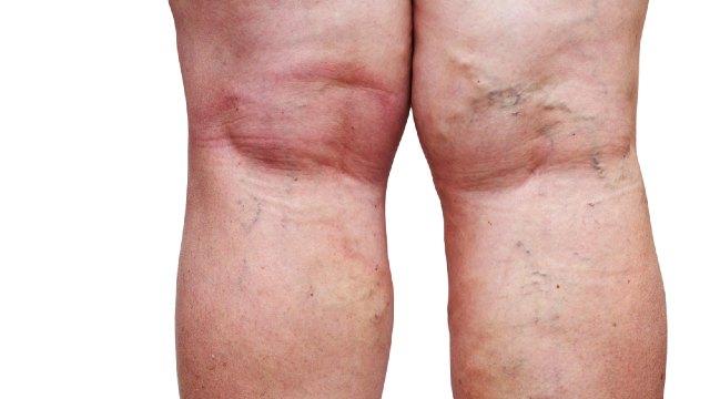
Reticular veins are the middle layer, and they are the ones most people have never heard of. They are larger than spider veins and sit slightly deeper, in the fat layer just beneath the skin rather than within it. Through the skin they read as blue or blue-green, sometimes with a faint greenish cast, and they form a flat net or lattice — reticular means net-like. They may be slightly palpable but they do not produce the ropy, raised contour of a varicose vein.
Their significance is functional rather than cosmetic, because a reticular vessel is often connected to the spider veins visible above it — a relationship covered in detail elsewhere in this library. What matters for telling the three types apart is simply that the middle layer is frequently the one doing the supplying, so it is not an isolated cosmetic finding. Ium Hazi Varicose Vein Clinic lists reticular veins alongside spider veins and small varicose veins among the vessels its cosmetic vein treatment addresses, and its stated method is to evaluate the visible veins first and, when necessary, use vascular ultrasound to establish whether underlying venous reflux or a deeper problem is present.
That sequence is the point. A vein you can barely see may be the reason a vein you can clearly see exists.
Varicose Veins
Varicose veins are the largest and most obvious of the three. They are enlarged, twisted superficial veins that bulge above the surface of the leg, with a ropy or knotted contour that can be felt easily and often becomes more prominent on standing. Ium Hazi Varicose Vein Clinic describes them as leg veins that have become enlarged or twisted where the venous valves are not working properly, allowing blood to pool or flow backward.
The important difference between varicose veins and the two smaller categories is that varicose veins are a recognized medical condition with a recognized natural history. MedlinePlus, the patient information service of the US National Library of Medicine, groups them under vascular diseases and describes them as swollen, twisted veins near the surface of the skin that can cause aching and heaviness. The National Heart, Lung, and Blood Institute (NHLBI) attributes them to weakened or damaged vein walls and valves that allow blood to collect in the leg rather than return efficiently.
The clinic lists swelling, heaviness, pain, visible veins, cramps, itching, and skin changes among the presenting complaints it sees, and states that varicose veins may be associated with pain, swelling, thrombosis, and skin ulcers. It also publishes the position that when varicose veins are left alone or permitted to advance, impaired circulation along with pain and swelling may follow, and that thrombosis or ulceration of the skin belongs to the more severe end of that range. No equivalent statement exists for spider veins in isolation, and that asymmetry is the practical difference between the categories.
What the Three Types Have in Common
All three are superficial veins, all three become visible because the vessel has widened, and all three can coexist in the same leg — frequently in the same square inch of skin. A leg can carry a bulging varicose vein behind the knee, a reticular lattice across the outer thigh, and a fan of spider veins at the ankle, all fed by the same failing valve higher up.
That is why treating them as three unrelated cosmetic issues tends to disappoint. Ium Hazi Varicose Vein Clinic's documented position is that a risk-reduction approach begins with an appropriate vascular assessment rather than treating visible veins on appearance alone, and that ultrasound is used to identify the affected veins and the reflux and to guide which treatment is selected.
Why "Cosmetic" and "Medical" Are Not Fixed Labels
Patients often want to know which category they are in, because they assume it determines whether the problem is serious. The honest answer is that the category is determined by what is found, not by what type of vein is visible.
Spider veins with healthy underlying deep veins and no reflux are genuinely a surface matter. The clinic states that ideal candidates for its surface laser treatment are people with small superficial spider veins or telangiectasias who have healthy underlying deep veins — the qualifier is doing real work in that sentence. Spider veins sitting above an incompetent saphenous trunk are a surface sign of a structural problem, and treating only what is visible leaves the cause untouched.
This runs in the other direction too. The clinic states that patients with deep venous insufficiency, active skin infections, recent tanning, or severe peripheral arterial disease are not suitable for laser treatment of spider veins, and that treatment may be postponed where there is an underlying venous problem requiring a different approach. The presence of a small vessel does not by itself qualify anyone for a small procedure.
The American Academy of Dermatology publishes patient guidance on safety in cosmetic treatment that translates directly to this situation: know who is performing the treatment, what their qualifications are, and what the plan is based on. Its list of questions to ask before a cosmetic procedure is a reasonable checklist for any vein consultation, including who will perform the treatment and what the realistic expectations are.
How Classification Drives Treatment Selection
The reason to name the vein type precisely is that the options available at each layer are different, and choosing between them is a clinical decision rather than a preference.
Vessels at the surface layer are addressed with energy delivered through the skin or with agents injected into the vessel, both of which work on small-caliber vessels and neither of which is designed to close a large trunk. The clinic documents surface laser treatment for spider veins and telangiectasias, and documents injection-based treatment as appropriate for selected spider veins, reticular veins, and certain small varicose veins, with the technique chosen according to the vein's size, location, depth, and the circulation behind it.
Larger refluxing trunks are a different proposition and are addressed with catheter-based closure or with surgical techniques. The clinic documents endovenous laser ablation, radiofrequency ablation, adhesive closure, mechanochemical ablation, point micro-surgery, and cryoablation among its options, and states that the appropriate choice depends on the vein involved, the severity of reflux, the symptoms, and the ultrasound findings. Its position is that having several methods available is what allows treatment to be matched to a patient's anatomy instead of one method being applied to every case.
Crucially, a treatment aimed at one layer is not a substitute for a treatment aimed at another. Closing surface vessels while a feeding trunk continues to leak is a known route to disappointment, and the clinic notes that new spider or varicose veins can develop even after successful treatment of existing veins.
What You Can and Cannot Establish From a Mirror
You can reasonably identify the caliber and the contour of what you see. Flat and thread-like, flat and net-like, or raised and ropy is a distinction most people can make accurately, and it is useful information to bring to an appointment.
What no visual inspection can establish is whether there is reflux behind any of it, which vein the reflux starts in, how long each episode of backward flow lasts, or whether the deep system is healthy. Those questions require dynamic imaging. The FDA explains that ultrasound imaging uses sound waves rather than ionizing radiation and can show internal structures in motion, including blood flowing through vessels, which is why it is the tool used for this rather than a photograph.
The practical implication is that the appropriate first step for visible leg veins of any size is a diagnostic consultation that includes vascular assessment, not a decision about which treatment you want. Documented results across vein types can be seen on the clinic's before and after page, and an assessment can be arranged through the contact page.
Who Would Classify Your Veins

Ium Hazi Varicose Vein Clinic works across the full range of vein calibers, from surface telangiectasias up to saphenous trunks, and has confined itself to venous disease since 2009 from premises in Seocho-gu, Seoul. Only one physician sees patients there. Under the clinic's single-director arrangement, Dr. Dong-Hyuk Kim handles each consultation, each diagnostic scan, and each procedure himself, so the classification of your veins and the treatment that follows are not handed between people.
His professional roles outside the clinic include Executive Director of the Korean Society of Phlebology and an adjunct professorship at Samsung Medical Center, and he is a founding member of the Venous Disease Research Society. His medical education was completed at Sungkyunkwan University School of Medicine. The practice publishes business registration 135-91-93104 and holds Registered Medical Institution for Attracting Foreign Patients certification granted under the Ministry of Health and Welfare. English-language support is available for international patients.
The Bottom Line
- Spider, reticular, and varicose veins describe three calibers of superficial vein at three depths: thread-like vessels within the skin, a flat blue net in the fat layer beneath it, and raised ropy veins that bulge above the surface.
- A clinician also uses a second, anatomical vocabulary — deep veins, saphenous trunks, subcutaneous veins, perforators — which answers a different question from the descriptive one.
- Reticular veins commonly feed clusters of spider veins, which is why the vessel causing a problem is often not the vessel a patient points at.
- Whether visible veins are a surface matter or a sign of structural disease depends on the ultrasound findings, not on which of the three types you have.
- Treatments are layer-specific, so establishing what feeds a visible vein before treating it is what keeps the plan coherent; a diagnostic consultation with vascular assessment is the appropriate starting point.
Frequently Asked Questions
How do I tell a reticular vein from a spider vein?
Size, color, and depth. Spider veins are finer, usually red or purple, sit within the skin, and are completely flat. Reticular veins are noticeably wider, read blue or blue-green through the skin because they lie in the fat layer beneath it, and often form a flat lattice. A reticular vein may also be slightly palpable, while a spider vein is not.
Can spider veins turn into varicose veins?
They are different vessels rather than different stages of the same vessel, so a spider vein does not grow into a varicose vein. What can happen is that both appear in the same leg because both are downstream of the same refluxing trunk. That is why the assessment looks for the source rather than cataloging the surface.
Do reticular veins need treating if they do not hurt?
That is decided at consultation rather than in advance. Ium Hazi Varicose Vein Clinic evaluates the visible veins and, where indicated, uses vascular ultrasound to determine whether underlying reflux or a deeper vein problem is present, then selects treatment according to the vein's characteristics. A vessel that feeds a visible cluster may be relevant to the plan even when it causes no symptoms itself.
Why did my spider veins come back after treatment?
Treated surface vessels are gradually broken down and absorbed, but the clinic states that new veins can develop over time, and that successfully treated veins generally do not reopen in the same way while venous disease itself can be progressive. Where a feeding vessel or an underlying refluxing trunk was never identified, new surface vessels can appear in the same area.
Which type of vein causes leg symptoms?
Aching, heaviness, swelling, cramps, itching, and skin changes are associated with varicose veins and with the venous reflux behind them, and Ium Hazi Varicose Vein Clinic lists exactly that cluster among the reasons patients seek assessment. Smaller vessels can accompany localized aching or leg tiredness, but persistent symptoms warrant an assessment of what lies deeper rather than treatment of the surface alone.
Does an ultrasound get done even for small veins?
The clinic's documented approach is to assess rather than to treat visible veins on appearance alone, using vascular ultrasound when it is needed to establish whether reflux or a deeper problem is present. The FDA notes that ultrasound imaging uses sound waves rather than ionizing radiation, which is part of why it can be used freely as an assessment tool.
This article is for general informational purposes and does not constitute individualized medical advice. Candidacy, technique, and expected outcomes can only be determined through a consultation with a qualified physician, and this article is not a self-diagnosis tool.
References
- MedlinePlus: Varicose Veins
- MedlinePlus: Vascular Diseases
- NHLBI: Varicose Veins
- FDA: Ultrasound Imaging
- AAD: Your safety
- AAD: Questions to ask before a cosmetic treatment
More Articles
Continue reading our clinical guides.
Will My Varicose Veins Come Back After Treatment?
A closed vein and a new varicose vein are different events. Here is what recurrence after vein treatment in Korea means and how follow-up imaging detects it.
Learn More

What to Pack Before Flying to Korea for Vein Surgery
What to pack before flying to South Korea for vein surgery: documents, medication and supplement lists, clothing, compression, and what to arrange at home.
Learn More
What Are Varicose Veins? Causes, Symptoms, and Signs
Varicose veins are enlarged, twisted leg veins caused by valve dysfunction. Learn the causes, the symptoms they produce, and when to see a vein specialist.
Learn More