Personalized Medical Care & Dedicated International Patient Concierge

Medical Guide
How Vascular Ultrasound Finds the Cause of Varicose Veins
Vascular ultrasound traces a visible varicose vein back to the failing valve that feeds it, identifying which vein leaks, where, and for how long.
Schedule a ConsultationHow Vascular Ultrasound Diagnoses the Root Cause of Varicose Veins
Medically reviewed by Dr. Dong-Hyuk Kim, Board-Certified General Surgeon, Medical Director of Ium Hazi Varicose Vein Clinic
Vascular ultrasound finds the root cause of varicose veins by following abnormal pressure backward through the leg until it reaches the vein that is actually leaking. The bulge you can see on your calf or thigh is usually the last link in a chain, not the first, and the link that matters for treatment often sits higher up and deeper down, where nothing is visible. Ium Hazi Varicose Vein Clinic in Seocho-gu, Seoul, uses this examination as the opening stage of a diagnostic consultation, and its findings are what a treatment plan is built from. This article explains how sound waves turn into a cause.
A Bulging Vein Is a Pressure Problem Arriving, Not Starting
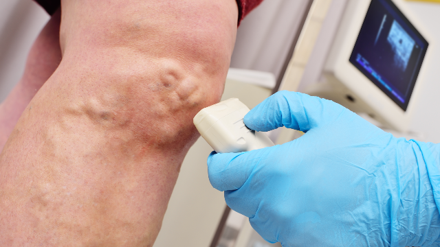
The vein you point to is rarely the vein that failed. Leg veins carry blood upward against gravity, and a series of one-way valves along the way stops the column from falling back between beats. The National Heart, Lung, and Blood Institute describes how a damaged or weakened valve allows blood to pool rather than continue toward the heart, raising pressure in the vessel segment below it.
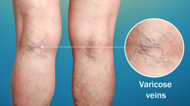
That raised pressure then travels. It passes from the failing trunk into the smaller branch veins connected to it, and those branches stretch under a load they were never built to carry. Over months and years the stretched branch becomes wide, twisted, and eventually visible through the skin. By the time you notice it, the wall of that branch has changed shape — but the valve that started the sequence may be fifteen or twenty centimeters away and completely invisible.
This is why an examination based on looking and touching reaches a limit quickly. MedlinePlus, the patient education service of the US National Library of Medicine, notes that enlarged, twisted leg veins commonly produce aching, heaviness, swelling, and night cramps, and that some people have visible veins with few complaints while others have marked symptoms. Neither the appearance nor the symptom load tells you which vessel is leaking. Two patients with an identical-looking bulge behind the knee can have entirely different sources feeding it, and the treatment that suits one would be aimed at the wrong target in the other.
The Questions the Scan Has to Answer
A scan earns the word "diagnostic" by answering specific questions rather than by producing a picture. Six of them matter most.
Which vein is refluxing? Ium Hazi Varicose Vein Clinic documents that its vascular ultrasound examination assesses the deep veins, the saphenous veins, the subcutaneous veins, and the perforating veins of the leg. Each group behaves differently, and naming the wrong one sends a treatment plan in the wrong direction.
Where along that vein does the backflow begin? A leak that starts at the top of a saphenous trunk and a leak that begins halfway down it are different anatomical problems, even though both can produce bulging veins near the ankle.
How far does the backflow extend? The length of the refluxing segment shapes how much vein needs to be addressed and by what means.
How long does each episode last? The examination timing findings are covered in the article on what the duplex scan measures; for root-cause purposes, what matters is that a timed finding is what turns a suspicion about a particular segment into a documented one.
Is the deep system working? The deep veins inside the muscle compartments handle most of the return from the leg, and their condition governs whether a superficial vein can be closed at all.
Which visible veins connect to the leaking segment? The surface veins you want gone have to be traced back to a source before anyone can say whether removing them alone would accomplish anything lasting.
Mapping Is a Route Description, Not a Photograph
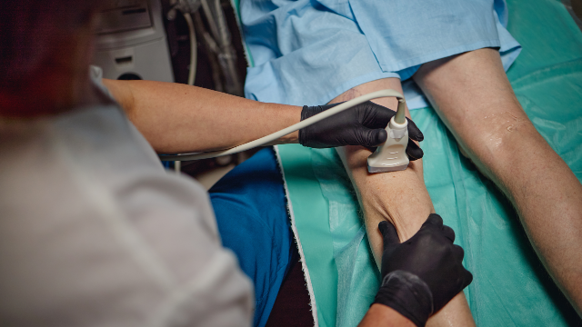
Venous mapping is the part of the examination that converts those answers into a plan of the leg. The physician follows each vessel along its course, records its size, shape, and position, and establishes which surface vein drains into which deeper one. What comes out of it is closer to a road map with the one-way signs marked than to a photograph of a leg.
That map is why treatment sequences at the clinic open the way they do. In its patient-facing description of endovenous laser ablation, the first step listed is ultrasound mapping of the affected vein, before any anesthesia or access. The same mapping step opens its endovenous vein ablation sequence. The device follows the map; the map does not follow the device.
Mapping also explains an outcome patients sometimes find surprising. A leg with a dozen visible veins may have one source, and a leg with two modest-looking veins may have several. The number of things you can see has no fixed relationship to the number of problems underneath.
Why the Examination Has to Be Watched, Not Photographed
Reflux is an event, not a state, so a frozen image cannot capture it. Blood in a refluxing vein flows correctly much of the time; what characterizes the disease is what happens in the moment when flow should stop and instead reverses. Catching that requires watching flow while it is changing.
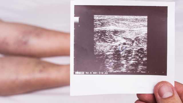
Ium Hazi Varicose Vein Clinic describes exactly this split in its own examination: the size, shape, and location of the veins are assessed from the images, while blood flow and reflux patterns are evaluated dynamically. The clinic states that its examination can demonstrate reflux in real time and that it explains findings using both ultrasound images and dynamic reflux flow, visually and audibly.
The US Food and Drug Administration explains that ultrasound imaging uses sound waves rather than ionizing radiation, and that one of its distinguishing capabilities is showing the movement of internal structures, including blood traveling through vessels, as it happens. That capability is not a convenience here. It is the entire reason the modality can diagnose a flow disorder at all.
What the Examination Rules Out, Which Matters as Much
Finding the cause also means excluding causes that would change the plan completely. Three exclusions carry real weight.
The first is a clot in the deep system. MedlinePlus describes deep vein thrombosis as a clot forming in a deep vein, usually in the lower leg or thigh, and notes that a fragment can break loose and travel to the lungs. The clinic lists active deep vein thrombosis among the conditions that can make a patient unsuitable for several of its treatments, so establishing the state of the deep veins is not optional background — it is a gate.
The second is arterial disease. The National Heart, Lung, and Blood Institute describes peripheral artery disease as narrowed arteries reducing blood flow to the limbs, often producing leg pain with walking. The clinic states that severe peripheral arterial disease may make someone an unsuitable candidate for endovenous ablation, and that people with significantly reduced arterial circulation may need alternative management rather than compression stockings. A leg with poor arterial inflow and a leg with venous reflux can both ache, and the wrong assumption leads to the wrong treatment.
The third is everything else that swells a leg. MedlinePlus notes that vascular disease covers a wide range of conditions affecting arteries, veins, and lymphatic vessels, and swelling alone does not identify which system is involved. An examination that finds no significant reflux is still a useful examination, because it redirects the search.
How the Cause Determines the Technique
Once the source is named, the technique follows from its physical characteristics, not from preference. The clinic's own candidacy criteria show the logic.
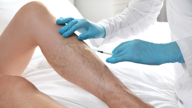
For endovenous laser ablation, the clinic states that suitability depends on ultrasound findings, vein anatomy, symptoms, medical history, and treatment goals — and that a vein too tortuous for safe fiber placement may not be suitable. A catheter or fiber has to travel inside the vessel, so a sharply winding vein is a mechanical constraint that no amount of enthusiasm overcomes.
For adhesive-based closure and for its other ablation methods, the clinic likewise describes candidacy being determined after examination and ultrasound evaluation, with the affected vein, anatomy, symptoms, and medical history all contributing. For point micro-surgery, the clinic's approach to prominent surface veins, candidacy follows clinical examination together with vascular ultrasound.
Dr. Dong-Hyuk Kim states his philosophy in these terms: identify accurately, through precise vascular ultrasound, what lies behind a patient's varicose veins, then provide only the treatment or combination of treatments that patient needs, leaving unnecessary procedures out. A principle like that is only operable if the cause has been established first. Without the scan, "only what is needed" has no reference point.
When the Cause Is Missed
The clinic is direct about the failure mode. It states that diagnosis and evaluation can sometimes miss or misinterpret the underlying cause, which can lead to delayed or inappropriate treatment, and that accurate vascular ultrasound assessment is important for understanding the condition and guiding the plan.
Two practical consequences follow. Treating a surface branch while leaving an upstream leak intact addresses the visible result while the pressure that produced it continues. And venous disease can be progressive: the clinic states plainly that new or untreated veins can develop problems in the future, so long-term results vary, and that a successfully treated vein does not prevent reflux appearing elsewhere later. No imaging study changes that, and no treatment removes the possibility of recurrence.
There are limits on the examination itself, stated honestly. Because nothing is injected or inserted, the clinic records no major contraindications to the study itself, while noting that it may have to be adjusted or deferred if the part of the leg in question cannot be reached properly. And a scan describes the veins at the moment it is performed; it does not forecast how the condition will behave over the following years.
Where the Scan Sits Inside the Visit
The examination is one component of a diagnostic consultation rather than a service to book on its own. The clinic describes the sequence as a medical history review, symptom assessment, physical examination where needed, diagnostic testing, review of the findings, and then a diagnosis with treatment recommendations. For varicose vein diagnosis and evaluation, it states that international patients generally need one day in Korea for the initial consultation, vascular ultrasound, and treatment planning, with the exact stay depending on what the findings show.
Findings are discussed during the appointment rather than issued later, and the clinic states that examination images can be saved to a USB drive on request. That record is worth having if you later see another physician.
One physician carries the whole sequence under the clinic's single-director system: the consultation, the diagnostic scan, any procedure, and the checks afterward. That physician is Dr. Dong-Hyuk Kim, Medical Director, board certified in general surgery, who completed his medical education at Sungkyunkwan University School of Medicine. His listed core specializations include vascular ultrasound diagnosis and evaluation alongside varicose vein diagnosis and treatment. More than 15 years dedicated to varicose vein care is what the practice states, and its own published statistic is 7,300+ saphenous vein surgeries performed 2009-2024. It operates under business registration 135-91-93104 and is a Registered Medical Institution for Attracting Foreign Patients certified by the Ministry of Health and Welfare, and it received the Medical Asia 2024 Grand Prize in the varicose vein category. You can read more about how the practice is organized on its about page, and arrange a diagnostic consultation through its contact page.
MedlinePlus publishes useful guidance on talking with your doctor, including writing your questions down beforehand and asking what the alternatives are. A consultation where backward flow is visible on a screen in front of you is a good setting for exactly those questions.
The Bottom Line
- A visible varicose vein is usually the downstream result of a valve failure elsewhere, so the vein you can see is often not the vein that needs treating.
- The scan answers specific questions: which vein refluxes, where the leak starts, how far it extends, how long each backflow episode lasts, whether the deep system is sound, and which surface veins connect to the source.
- Reflux has to be observed while flow changes, which is why the clinic evaluates blood flow dynamically rather than from still images alone.
- Ruling conditions out matters as much as ruling them in: deep vein thrombosis and severe peripheral arterial disease both change or exclude treatment options.
- Vein anatomy found on the scan determines which technique is mechanically possible, and venous disease can still progress or recur after successful treatment.
Frequently Asked Questions
Why can't a physician diagnose varicose veins by looking at my legs?
Looking identifies which veins have enlarged, but not why. The failing valve driving the problem is frequently in a deeper or higher vein that produces no visible sign at all. Vascular ultrasound is what connects the surface finding to its source.
What does "reflux" actually mean on an ultrasound report?
Reflux means blood traveling backward in a vein instead of continuing toward the heart. What the clinic's examination records about that backward flow is described in the article devoted to the duplex scan itself; this article is concerned with the next question, which is which vein the reflux originates in.
Which veins are checked during the examination?
The clinic documents assessment of the deep veins, the saphenous veins, the subcutaneous veins, and the perforating veins of the leg. Each group contributes different information, and the deep veins in particular influence whether superficial treatment is appropriate.
Can the scan show a blood clot?
Evaluating the deep venous system is part of the examination, and MedlinePlus describes deep vein thrombosis as a clot in a deep vein, most often in the leg. The clinic lists active deep vein thrombosis among the conditions that can make several of its treatments unsuitable, so this finding directly affects what can be offered.
Does the ultrasound decide which treatment I get?
The findings determine which techniques are mechanically and medically possible, and the physician then discusses options within that set. The clinic states that suitability for its ablation treatments depends on ultrasound findings, vein anatomy, symptoms, medical history, and treatment goals.
Is there any radiation or injection involved?
No. The FDA explains that ultrasound imaging produces its pictures using sound waves rather than ionizing radiation. The clinic describes the study as non-invasive, generally requiring no anesthesia, with mild pressure from the probe as the usual extent of any discomfort.
This article is for general informational purposes and does not constitute individualized medical advice. Candidacy, technique, and expected outcomes can only be determined through a consultation with a qualified physician, and this article is not a self-diagnosis tool.
References
- NHLBI: Varicose Veins
- MedlinePlus: Varicose Veins
- FDA: Ultrasound Imaging
- MedlinePlus: Deep Vein Thrombosis
- NHLBI: Peripheral Artery Disease
- MedlinePlus: Vascular Diseases
- MedlinePlus: Talking With Your Doctor
More Articles
Continue reading our clinical guides.
Will My Varicose Veins Come Back After Treatment?
A closed vein and a new varicose vein are different events. Here is what recurrence after vein treatment in Korea means and how follow-up imaging detects it.
Learn More

What to Pack Before Flying to Korea for Vein Surgery
What to pack before flying to South Korea for vein surgery: documents, medication and supplement lists, clothing, compression, and what to arrange at home.
Learn More
What Are Varicose Veins? Causes, Symptoms, and Signs
Varicose veins are enlarged, twisted leg veins caused by valve dysfunction. Learn the causes, the symptoms they produce, and when to see a vein specialist.
Learn More