Personalized Medical Care & Dedicated International Patient Concierge

Medical Guide
Duplex Ultrasound: First Step in Diagnosing Venous Reflux
A vascular duplex ultrasound examines the deep, saphenous, subcutaneous and perforating veins and shows reflux in real time, guiding the treatment choice.
Schedule a ConsultationVascular Duplex Ultrasound Examination: The First Step in Diagnosing Venous Reflux
Medically reviewed by Dr. Dong-Hyuk Kim, Board-Certified General Surgeon, Medical Director of Ium Hazi Varicose Vein Clinic
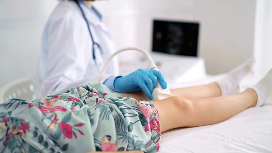
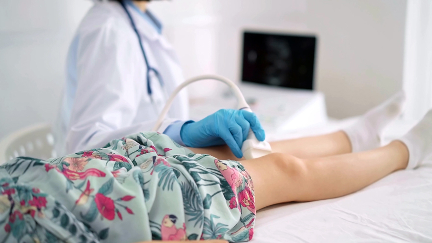
A vascular duplex ultrasound examination evaluates the condition of the leg veins, including the deep veins, the saphenous veins, the subcutaneous veins, and the perforating veins. It assesses the size, shape, and location of each vein and evaluates blood flow, and it can demonstrate reflux, meaning blood flowing backward, as it happens. At Ium Hazi Varicose Vein Clinic in Seocho-gu, Seoul, this examination is the opening step of a diagnostic consultation, and the findings are what determine which treatment, if any, is appropriate. This article explains what is examined, how the findings are shown to you, and why the diagnosis drives everything that follows.
What Venous Reflux Is and Why It Needs Imaging
Reflux is the term for blood traveling the wrong way inside a vein rather than heading steadily back toward the heart. One-way valves sit along the leg veins to prevent exactly that between heartbeats. According to the National Heart, Lung, and Blood Institute (NHLBI), a valve that has weakened or been damaged lets blood fall back and collect below it, so pressure climbs in the vessels underneath.
The difficulty is that reflux is invisible from outside the leg. MedlinePlus, the patient-education service of the US National Library of Medicine, lists aching, heaviness, cramping, swelling, and skin changes among what enlarged, twisted leg veins can produce. What you can see is the consequence. Where the leak starts, which vein is involved, how far the backward flow extends, and how long each episode of backflow lasts are all questions that a visual examination cannot answer.
This is why the examination is diagnostic rather than cosmetic. Two legs that look similar can have entirely different problems underneath, and the treatment appropriate for one would be the wrong choice for the other.
What "Duplex" Means
Duplex refers to combining two kinds of ultrasound information in one examination. The first is a grayscale image showing the anatomy: where the vein is, how deep it sits, how wide it is, and what shape it has. The second is Doppler information about movement, showing the direction and speed of blood flowing inside that vein. Putting the two together lets the examiner say not only that a vein is enlarged but that blood inside it is traveling the wrong way.
The US Food and Drug Administration (FDA) explains that ultrasound imaging uses high-frequency sound waves to produce pictures of structures inside the body, that it involves no ionizing radiation, and that it can show internal structures in motion, including blood flowing through vessels. The FDA also notes that ultrasound is generally regarded as safe when performed appropriately by trained operators, which is why it is the standard imaging approach for this problem.
Ium Hazi Varicose Vein Clinic describes its examination as a Doppler assessment of the deep, saphenous, subcutaneous, and perforator veins of the leg, identifying the size, shape, and location of the veins along with the direction and duration of reflux. Duration matters as much as direction: a brief backward movement as a valve closes is not the same finding as sustained backflow.
What Is Actually Examined
Four groups of veins are assessed, and each one tells the physician something different.
The deep veins run inside the muscle compartments and carry most of the blood returning from the leg. Their condition affects which superficial treatments are safe to consider at all, and a clot or obstruction there changes the entire plan. MedlinePlus notes that deep vein thrombosis is a clot in one of these deep veins, most often in the leg, and that part of a clot can travel to the lungs.
The saphenous veins are the long superficial trunks running up the leg, and they are a common origin point for reflux that later shows up as visible bulging veins lower down.
The subcutaneous veins are the smaller superficial vessels beneath the skin, including the ones patients can see and point to.
The perforating veins connect the superficial system to the deep system through the muscle layer. When a perforator's valve fails, high pressure from the deep system is transmitted outward to the superficial veins, which can keep a surface problem going even after other veins have been addressed.
The clinic states that the size, shape, and location of the veins are assessed visually on the images, while blood flow and reflux patterns are evaluated dynamically, meaning while flow is changing rather than from a frozen picture.
How the Examination Is Performed
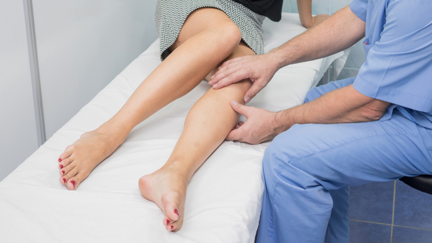
The sequence is short and non-invasive. The examination area is prepared for ultrasound. A probe is moved over the leg to examine the veins. The deep, saphenous, subcutaneous, and perforating veins are each assessed. Blood flow and reflux are observed in real time. The findings are then explained using both the ultrasound images and the dynamic reflux flow. If requested, the images can be saved directly to a USB drive.
No anesthesia is required, because this is a non-invasive diagnostic examination, and most patients feel little or no discomfort. The examination is generally completed during a single clinic visit, with the exact time depending on how many vein areas need to be assessed. Usually no special preparation is needed for a leg vascular ultrasound, though you should follow any instructions the clinic gives you beforehand.
Afterward there is nothing to recover from. No specific recovery period is required and normal activities can generally resume immediately, unless another procedure is performed at the same visit. No special aftercare is needed.
Findings Are Shown to You While They Happen
One feature of the clinic's documented approach deserves attention, because it changes the character of the appointment. The results are available during the examination itself: the clinic evaluates and explains the venous condition using real-time ultrasound images and dynamic reflux flow, visually and audibly. You are not waiting days for a report.
The clinic explains reflux using real-time dynamic ultrasound rather than static images alone, and patients can request that their examination images be saved directly to a USB drive. That means you leave with a record of your own study rather than only with a verbal summary, which is useful if you later consult another physician or need continuity of care after returning home.
Seeing the backflow on screen also makes the treatment discussion concrete. When a physician points to blood moving the wrong way in a specific named vein, the recommendation that follows stops being an assertion and becomes something you can ask direct questions about. MedlinePlus publishes practical guidance on talking with your doctor that fits this moment well: prepare your questions in advance, ask what the alternatives are, and ask what happens if nothing is done. Its guidance on evaluating health information is equally relevant, because what you see on the screen with your physician is a better basis for a decision than a claim on a marketing page.
Why the Diagnosis Drives the Treatment Choice
The examination is not an end in itself. It exists because the treatment decision depends entirely on what is found, and the clinic's documented approach reflects that at every step.
Dr. Dong-Hyuk Kim states his philosophy as establishing accurately, by precise vascular ultrasound, what lies behind a patient's varicose veins, and then delivering nothing beyond the treatment or combination of treatments that patient needs, with unnecessary procedures left out. That principle only works if the underlying cause has actually been identified first.
The pattern repeats across the clinic's services. Candidacy for endovenous ablation depends on ultrasound findings, vein anatomy, symptoms, medical history, and treatment goals. Suitability for adhesive-based closure is determined through vascular assessment and imaging. Point micro-surgery candidacy follows clinical examination and vascular ultrasound. Whether sclerotherapy is appropriate, and which veins should be treated, may be determined by a vascular examination and duplex ultrasound. Even for compression therapy, vascular assessment such as Doppler or duplex ultrasound may be performed where clinically indicated. Non-surgical vein care options depend on the vein involved, the severity of reflux, the symptoms, and the ultrasound findings.
In other words, every path forward passes through the same gate. A diagnostic consultation with vascular assessment is the appropriate first step for anyone with leg symptoms or visible veins, and it is the step that makes an individualized plan possible rather than a generic one. You can arrange that consultation through the clinic's contact page.
What the Examination Cannot Tell You
Honesty about limits belongs here. A duplex study describes the state of the veins at the moment it is performed. It does not predict how venous disease will progress, and venous disease can be progressive. It does not by itself determine that any particular treatment will succeed, and outcomes vary by patient, anatomy, and technique.
There are also practical limits to the examination. The clinic notes that there are generally no major contraindications because the study is non-invasive, but that it may need to be modified or postponed when the examination area cannot be adequately accessed. Skin conditions, dressings, wounds, or swelling in the area being scanned can affect what is obtainable.
Risks are minimal but not worth overstating in either direction. Vascular ultrasound is generally considered a safe, non-invasive examination, significant complications are uncommon, and there is no radiation exposure. Mild pressure or temporary discomfort from the probe may occur, particularly over tender areas.
Follow-Up Imaging After Treatment
Ultrasound is used again after treatment, not only before it. The clinic states that the examination helps visualize abnormal backflow and compare blood-flow findings before and after treatment. Its aftercare after a procedure is built around scheduled vascular ultrasound examinations together with individualized advice on medical compression stockings, and a next-day check may be recommended where necessary.
This matters for how a result is judged. Appearance is an unreliable measure on its own, because bruising and swelling take weeks to settle and because a closed vein is not always invisible. Imaging shows whether the treated vein has actually closed and what the flow pattern looks like now. It also picks up new reflux in a different vein, which can develop later even after a successful treatment.
The clinic's quality-control process includes doctor-led diagnosis, consultation, surgery, and post-operative care, pre-operative vascular ultrasound and clinical evaluation, a deliberately limited caseload, strict sterilization, single-use devices that are not reused, and post-operative ultrasound monitoring and follow-up care.
Who Performs the Examination
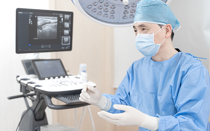
Under the clinic's single-director system, the consultation, the diagnostic scan, any procedure that follows, and the later checks are all carried out by the same physician. Dr. Dong-Hyuk Kim serves as Medical Director, is board certified in general surgery, and received his medical education at Sungkyunkwan University School of Medicine. His listed core specializations include vascular ultrasound diagnosis and evaluation, varicose vein diagnosis and treatment, point micro-surgery, radiofrequency ablation, and fourth-generation 1940 nm endovenous laser surgery.
The practice states over 15 years given to varicose vein care, and its own published statistic is 7,300+ saphenous vein surgeries between 2009 and 2024. Documented equipment includes a Samsung Medison V8 ultrasound system and a VeinViewer Flex vein-imaging device. It operates under business registration 135-91-93104 and is recognized by the Ministry of Health and Welfare under the designation Registered Medical Institution for Attracting Foreign Patients. It won the varicose vein category of the Medical Asia 2024 Grand Prize. English-language support is offered to international patients, and more about the practice is on the clinic's about page.
MedlinePlus offers general guidance on choosing a doctor or health care service, including asking about credentials and about how much experience the clinician has with your particular condition. Continuity between the person who scans your leg and the person who treats it is a reasonable thing to ask about anywhere.
The Bottom Line
- A vascular duplex ultrasound assesses the deep, saphenous, subcutaneous, and perforating veins, and evaluates the size, shape, and location of each along with blood flow.
- The study shows reflux in real time, including its direction and duration, which a physical examination cannot reveal.
- Findings are explained during the examination using both images and dynamic flow, and the images can be saved to a USB drive on request.
- The examination is non-invasive, needs no anesthesia, requires no recovery, and carries no radiation exposure; mild probe pressure is the usual extent of discomfort.
- The diagnosis is what determines which treatment is appropriate, so a diagnostic consultation with vascular assessment is the first step rather than a formality.
Frequently Asked Questions
What does a vascular duplex ultrasound examine?
It evaluates the leg veins, including the deep veins, the saphenous veins, the subcutaneous veins, and the perforating veins. The size, shape, and location of the veins are assessed on the images, while blood flow and reflux are evaluated dynamically. The clinic identifies both the direction and the duration of reflux.
Does the examination hurt?
Vascular ultrasound is generally painless. Some patients notice mild pressure or temporary discomfort as the probe is moved across the skin, particularly over a tender area. No anesthesia is required because the study is non-invasive.
How long does it take, and do I need more than one visit?
The examination is generally completed during a single clinic visit. The exact duration depends on the number and areas of veins being evaluated. International patients should allow additional time if a consultation, further testing, or treatment is planned in the same trip.
When do I find out the results?
During the examination. The clinic evaluates and explains the venous condition using real-time ultrasound images and dynamic reflux flow, so the findings are discussed as they are obtained rather than issued later as a report.
Can I get a copy of my own images?
Yes. The clinic states that examination images can be saved directly to a USB drive when requested. Having your own study is useful for continuity of care if you consult another physician after returning home.
Is there any radiation involved?
No. The FDA explains that ultrasound imaging uses sound waves rather than ionizing radiation and is generally considered safe when used appropriately by trained operators. That is one reason it is suitable for repeated use, including follow-up scans after treatment.
Why is imaging repeated after treatment?
Because appearance alone is an unreliable measure of what happened inside the vein. The clinic compares blood-flow findings before and after treatment, and its post-operative aftercare includes scheduled vascular ultrasound examinations. Follow-up imaging also detects new reflux in a different vein, which can develop over time.
Do I need to prepare for the examination?
Usually no special preparation is required for a leg vascular ultrasound. Follow any specific instructions the clinic gives you beforehand, and bring relevant medical records, previous test results, and a list of your current medications and symptoms.
This article is for general informational purposes and does not constitute individualized medical advice. Candidacy, technique, and expected outcomes can only be determined through a consultation with a qualified physician, and this article is not a self-diagnosis tool.
References
- FDA: Ultrasound Imaging
- MedlinePlus: Varicose Veins
- MedlinePlus: Deep Vein Thrombosis
- MedlinePlus: Vascular Diseases
- MedlinePlus: Talking With Your Doctor
- MedlinePlus: Evaluating Health Information
- MedlinePlus: Choosing a Doctor or Health Care Service
- NHLBI: Varicose Veins
- CDC: About Venous Thromboembolism
More Articles
Continue reading our clinical guides.
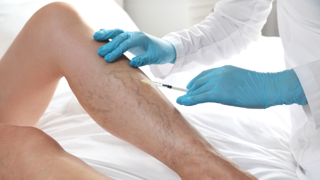
Will My Varicose Veins Come Back After Treatment?
A closed vein and a new varicose vein are different events. Here is what recurrence after vein treatment in Korea means and how follow-up imaging detects it.
Learn More

What to Pack Before Flying to Korea for Vein Surgery
What to pack before flying to South Korea for vein surgery: documents, medication and supplement lists, clothing, compression, and what to arrange at home.
Learn More
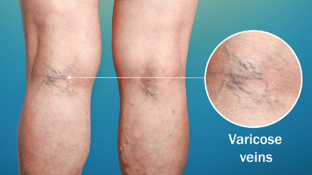
What Are Varicose Veins? Causes, Symptoms, and Signs
Varicose veins are enlarged, twisted leg veins caused by valve dysfunction. Learn the causes, the symptoms they produce, and when to see a vein specialist.
Learn More